Scanning Electron Images of a Cerambycid Beetle
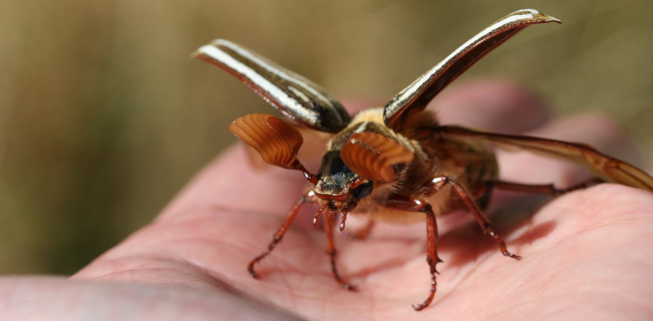
Yesterday I had the extreme good fortune to be able to use the scanning electron microscope (SEM) at University of Washington’s Friday Harbor Labs (San Juan Island). We put a Cerambycid beetle under the SEM and “WOW,” the photos were phenomenal! Here’s a few for you to see. Below is a photo of the beetle’s compound eye. Just think of all the information each of those facets receives and processes.

Cerambycid beetle compound eye, imaged under scanning electron microscope at Friday Harbor Labs, San Juan Island, WA
Next, you see an image of the beetle head. It shows the antennal insertion points, the compound eyes, frons, clypeus, labrum, mandibles, and bristly setae.
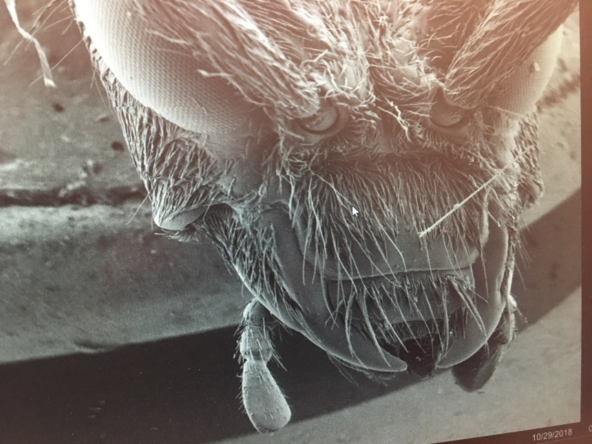
SEM anterior, dorsal view of cerambycid head.
If you’re interested in learning more about the morphological features, here’s a pretty good diagram below for reference.

image from http://www.faculty.ucr.edu/~legneref/biotact/bc-51b.htm
The last image for you is of the beetle’s tarsi (the foot). This is an important identification feature for many insects. Imagine that! When I was working on my masters degree from the University of Florida, I had an amazing taxonomy professor who was an expert on Coleoptera (the beetles). He created identification keys for Florida beetles and you can take a look at them here: http://www.entnemdept.ufl.edu/choate/beetles.pdf  Well, I’m looking forward to using the SEM again and my next imaging will hopefully include the sponging mouthparts of a fly. Stay tuned!
